Understanding bile duct anatomy is crucial for successful ERCP procedures
Anatomical variations exist. Be familiar with the following classifications for successful procedures:
Here’s a quick overview:
Left hepatic duct: Drains segments II, III, and IV.
Right hepatic duct: Shorter and drains the right posterior sectoral duct (VI/VII) and right anterior sectoral duct (V/VIII).
Caudate lobe drainage:
78% of cases: Into the right and left hepatic ducts.
15% of cases: By the left hepatic ductal system only.
7% of cases: Into the right hepatic system.
Anatomical variations exist. Be familiar with the following classifications for successful procedures:
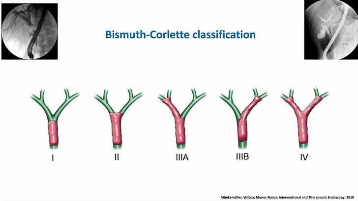
Bismuth-Corlette: Classifies bile duct strictures.
Strasberg: Classifies bile duct injuries following cholecystectomy.
Du Bois: Classifies bile duct injury after liver transplant.
The appearance of the papilla can also influence cannulation difficulty. Study papilla morphology and classifications (like the Haraldson classification shown here) to anticipate cannulation challenges.
Visit EndoCollab for additional resources, images, and videos to further enhance your understanding of this important topic!


